Eye Care Services & Treatment
Providing comprehensive eye care solutions for all your vision needs and concerns.


OPD
Refraction
Intra Ocular Pressure (IOP) Measurement
Retinal Examination (Fundoscopy)
A scan biometry
Lacrimal sac syringing
Surgical
Cataract Surgery -
Phacoemulsification Cataract Surgery
Microincision Phacoemulsification Cataract Surgery
Small Incision Cataract surgery
Glaucoma Surgery -
Trabeculectomy
Trabeculectomy with MitoMycin C (MMC)
Pterygium Surgery -
Pterygium excision with conjunctival graft with Bandage Contact Lens
Pterygium excision with conjunctival graft No Sutures No Glue Technique
Lacrimal Sac Surgery -
Dacryocystectomy (DCT) surgery
Laser
Refractive -
LASIK
PRK
NdYAG Laser Capsulotomy for Posterior Capsular Opacity (PCO)
Glaucoma - Laser Peripheral Iridotomy
- Brief description of each service -
OPD -
Refraction -
This is done to calculate the power of glasses. Instrument used is Autorefractokeratometer.
Wearing correct glasses for distance, near and intermediate distance reduces strain on eyes.
This should be done once in a year.
Intra Ocular Pressure (IOP) Measurement -
IOP is pressure within the eye.
Normal range is 10 to 21 mm of Hg.
IOP is measured with the help of Tonometer.
Persistent readings more than 21 mm of Hg are suspicious of Glaucoma.
Further detailed examination is necessary in these cases.
A Scan Biometry -
This is done to calculate the power of intraocular lens in patient undergoing cataract surgery.
Instrument used is A Scan Biometer.
Lacrimal sac syringing -
This is done in patient with persistent watering.
It access the patency of lacrical sac & drainage passage.
Surgical & Laser -
Surgical -
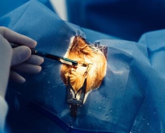
Cataract Surgery -
Phacoemulsification Cataract Surgery - In this procedure 2.8 mm incision is made to remove the cataract lens. Cataract lens removed by ultrasonic energy.
Foldable intra ocular lens of appropriate type & power is put inside the bag.
2.8 nm incision gives early visual recovery.
Microincision Phacoemulsification Cataract Surgery -
In this surgery incision is 1.8 to 2 mm.
Small Incision Cataract Surgery -
Incision size s around 5.5 to 6 mm. Here the cataract lens is removed manually. Incision is 2 mm away from the Cornea. So the result are comparable with Phacoemulsification Cataract Surgery.
This procedure is indicated in hard brown black cataract, comparamined corneas.
Glaucoma Surgery
Trabeculectomy -
This procedure is indicated in Glaucoma not controlled with the medical treatment.
In this surgery, a small passage is made within the inner coat of the eye ball. So the intra ocular fluid passes through the passage to outside of eye ball. This reduces the intra ocular pressure (IOP).
Trabeculectomy with MitoMycin C (MMC) -
Indicated in more advance form of Glaucoma. Here MMC drug is used before making the passage.
Pterygium Surgery -
Pterygium excision with autollogus conjunctival graft with Bandage Contact Lens.
In this procedure, pterygium tissue is dissected & removed. Conjunctival graft is placed over the defect. Bandage contact lens is put to keep the graft in place.
Pterygium excision with autollogus conjunctival graft No Sutures No Glue Technique.
In this procedure sutures or glue is not used. Graft is placed over the defect for about 10 to 15 minutes. So it gets attached by its own.
Incision & Drainage (I&D) of Chalazion -
Chalazion is the cyst which contain oily sebaceous material. It occurs in the eyelids.
Under local anesthesia, incision is made either inside or outside of lid and the oily contents are drained. The eye is patched for 12 hrs. after the procedure to avoid bleeding from the incision site.
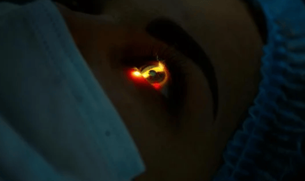
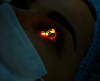
Laser -
Refractive Procedure - LASIK and PRK
"Don't Shadow Your World Behind Glasses"
These procedures are done for correction of refractive error particularly Myopia.
These procedures are done with Nidek EC 5000.
Refractive Laser System -
After basic eye exanimation that includes corneal thickness, appointment is given for LASIK.
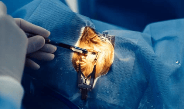
How LASIK is done?
The Flap is created - After refractive error calculation, an extremely thin flap is created at the cornea. This step is performed by Microkeratome. The flap is folded back like a page of book.
The vision defect is corrected with excimer laser.
The laser sculpts the corneal tissue within few seconds to an exact curvature calculated to correct the visual error.
The flap forms body's sticking plaster. Finally the flap is folded back to its original position as protective layer.
The entire procedure is painless, takes last couple of minutes and is done under topical anaesthesia [Only drops are put in the eye to numb the cornea].
Visual recovery is rapid and there is little or no past operative pain. Eye drops are given to be put for four weeks.
The entire procedure is done at - Akash Laser Center, Pune.
NdYAG Laser Capsulotomy for PCO -
This is OPD procedure. PCO i.e posterior capsular opacification develops few years after the cataract surgery. It a body's response in which few lens cells grow over the posterior capsule over which the intra ocular lens (IOL) is fitted.
In this procedure the opacity is dissolved with NdYAG laser. Entire procedure is done under topical anesthesia i.e drops are put in eye to numb it. The procedure is over in 2 to 3 minutes.
After the procedure, drops are given to put in eye for 10 days.
Laser Peripheral Iridotomy -
This procedure done in certain cases of Glaucoma as treatment or prophylaxis.In this, a small opening is made between anterior and posterior compartments of eye with the help of NdYAG laser.
This passage allows free flow of ocular fluid between two compartments and thus reduces intra ocular pressure(IOP)
It is done under tropical anaesthesia [i.e drops are put in eye to numb it]. The entire procedure takes 5 minutes.
Drops are given to be put in eye for about 14 days.
Equipments -
OPD -
Autorefractokeratometer Huvitz and Topcon
Slit Lamp - LABOMED
Tonometer
Lensometer
Vision Chart
Color Vision Chart
Gonioscope
90 D Fundoscopy Volk Lens Wide Angle
Laser -
NdYAG Laser
Surgical -
Opticon Phacoemulsification System
Zeiss IFR Microscope With Omniglow Retroillumination System
LASIK -
Nidek EC 5000 Refractive Laser System@AkashLaserCenter
Consultant Eye Surgeon at -
Moraya Multispeciality Hospital Chinchwad.
Gandhi Nursing Home, Nigdi
Akash Laser Center, Pune
MIMER Medical College,Talegaon Dabhade
Appointment Booking Details -
Walking only.
For any query -
WhatsApp on - 9623138991
Lacrimal Sac syringing -
Dacryocystectomy (DCT) surgery
This procedure is indicated in Chronic Dacryocystitis in old age.
The Lacrimal Sac is removed.
Patient may complain of watering after this procedure.
Retinal Examination (Fundoscopy) -
This evaluates the status of retina.
This is done with 90 D lens on slit lamp OR with Ophthalmoscope.
Fundoscopy is particularly important in Diabetic patients.
Depending on the findings, drops/ laser/ intravitreal injection can be given.
This will prevent the progression of vision loss.
- Brief description of each service -
OPD -
Refraction -
This is done to calculate the power of glasses. Instrument used is Autorefractokeratometer.
Wearing correct glasses for distance, near and intermediate distance reduces strain on eyes.
This should be done once in a year.
Intra Ocular Pressure (IOP) Measurement -
IOP is pressure within the eye.
Normal range is 10 to 21 mm of Hg.
IOP is measured with the help of Tonometer.
Persistent readings more than 21 mm of Hg are suspicious of Glaucoma.
Further detailed examination is necessary in these cases.
A Scan Biometry -
This is done to calculate the power of intraocular lens in patient undergoing cataract surgery.
Instrument used is A Scan Biometer.
Lacrimal sac syringing -
This is done in patient with persistent watering.
It access the patency of lacrical sac & drainage passage.
Surgical & Laser -
Cataract Surgery -
Phacoemulsification Cataract Surgery - In this procedure 2.8 mm incision is made to remove the cataract lens. Cataract lens removed by ultrasonic energy.
Foldable intra ocular lens of appropriate type & power is put inside the bag.
2.8 nm incision gives early visual recovery.
Microincision Phacoemulsification Cataract Surgery -
In this surgery incision is 1.8 to 2 mm.
Small Incision Cataract Surgery -
Incision size s around 5.5 to 6 mm. Here the cataract lens is removed manually. Incision is 2 mm away from the Cornea. So the result are comparable with Phacoemulsification Cataract Surgery.
This procedure is indicated in hard brown black cataract, comparamined corneas.
Trabeculectomy with MitoMycin C (MMC) -
Indicated in more advance form of Glaucoma. Here MMC drug is used before making the passage.
Pterygium Surgery -
Pterygium excision with autollogus conjunctival graft with Bandage Contact Lens.
In this procedure, pterygium tissue is dissected & removed. Conjunctival graft is placed over the defect. Bandage contact lens is put to keep the graft in place.
Pterygium excision with autollogus conjunctival graft No Sutures No Glue Technique.
In this procedure sutures or glue is not used. Graft is placed over the defect for about 10 to 15 minutes. So it gets attached by its own.
Incision & Drainage (I&D) of Chalazion -
Chalazion is the cyst which contain oily sebaceous material. It occurs in the eyelids.
Under local anesthesia, incision is made either inside or outside of lid and the oily contents are drained. The eye is patched for 12 hrs. after the procedure to avoid bleeding from the incision site.
Laser -
Refractive Procedure - LASIK and PRK
"Don't Shadow Your World Behind Glasses"
These procedures are done for correction of refractive error particularly Myopia.
These procedures are done with Nidek EC 5000.
Refractive Laser System -
After basic eye exanimation that includes corneal thickness, appointment is given for LASIK.
NdYAG Laser Capsulotomy for PCO -
This is OPD procedure. PCO i.e posterior capsular opacification develops few years after the cataract surgery. It a body's response in which few lens cells grow over the posterior capsule over which the intra ocular lens (IOL) is fitted.
In this procedure the opacity is dissolved with NdYAG laser. Entire procedure is done under topical anesthesia i.e drops are put in eye to numb it. The procedure is over in 2 to 3 minutes.
After the procedure, drops are given to put in eye for 10 days.
Laser Peripheral Iridotomy -
This procedure done in certain cases of Glaucoma as treatment or prophylaxis.In this, a small opening is made between anterior and posterior compartments of eye with the help of NdYAG laser.
This passage allows free flow of ocular fluid between two compartments and thus reduces intra ocular pressure(IOP)
It is done under tropical anaesthesia [i.e drops are put in eye to numb it]. The entire procedure takes 5 minutes.
Drops are given to be put in eye for about 14 days.
Equipments -
OPD -
Autorefracto Keratometer Huvitz and Topcon
Slit Lamp - LABOMED
Tonometer
Lensometer
Vision Chart
Color Vision Chart
Gonioscope
90 D Fundoscopy Volk Lens Wide Angle
Laser -
NdYAG Laser
Surgical -
Opticon Phacoemulsification System
Zeiss IFR Microscope With Omniglow Retroillumination System
LASIK -
Nidek EC 5000 Refractive Laser System@AkashLaserCenter
Consultant Eye Surgeon at -
Moraya Multispeciality Hospital Chinchwad.
Gandhi Nursing Home, Nigdi
Akash Laser Center, Pune
MIMER Medical College,Talegaon Dabhade
Appointment Booking Details -
Walking only.
For any query -
WhatsApp on - 9623138991
Lacrimal Sac syringing -
Dacryocystectomy (DCT) surgery
This procedure is indicated in Chronic Dacryocystitis in old age.
The Lacrimal Sac is removed.
Patient may complain of watering after this procedure.
Retinal Examination (Fundoscopy) -
This evaluates the status of retina.
This is done with 90 D lens on slit lamp OR with Ophthalmoscope.
Fundoscopy is particularly important in Diabetic patients.
Depending on the findings, drops/ laser/ intravitreal injection can be given.
This will prevent the progression of vision loss.
Glaucoma Surgery
Trabeculectomy -
This procedure is indicated in Glaucoma not controlled with the medical treatment.
In this surgery, a small passage is made within the inner coat of the eye ball. So the intra ocular fluid passes through the passage to outside of eye ball. This reduces the intra ocular pressure (IOP).
Surgical -
How LASIK is done?
The Flap is created - After refractive error calculation, an extremely thin flap is created at the cornea. This step is performed by Microkeratome. The flap is folded back like a page of book.
The vision defect is corrected with excimer laser.
The laser sculpts the corneal tissue within few seconds to an exact curvature calculated to correct the visual error.
The flap forms body's sticking plaster. Finally the flap is folded back to its original position as protective layer.
The entire procedure is painless, takes last couple of minutes and is done under topical anaesthesia [Only drops are put in the eye to numb the cornea].
Visual recovery is rapid and there is little or no past operative pain. Eye drops are given to be put for four weeks.
The entire procedure is done at - Akash Laser Center, Pune.
Client Feedback
Our patients appreciate our dedicated and professional eye care services.
The staff at Shree Netralaya were incredibly helpful and attentive during my visit.
John Doe
Mumbai
I received exceptional care at Shree Netralaya. The team was knowledgeable and made me feel comfortable throughout my appointment. Highly recommend their services for eye care.
Jane Smith
Delhi
★★★★★
★★★★★
Care
Providing expert eye care for your vision needs.
Contact
Health
- 9422081910
© 2025. All rights reserved.
- 9623138991
-